A modular, open CT system
with freely interchangeable sensor modules
Commercially available CT scanners offer little scope for adding extra components or connecting
custom sensors – which significantly hampers the corresponding research and development efforts.
The overall Kids-CT project therefore pursued the vision of a unique, modular medical scanner
system distinguished by a variety of freely interchangeable sensor modules for image
acquisition – with a CT device as the base module and open hardware and software interfaces.
The subproject carried out by Dornheim focused on multimodality: the goal of deriving a higher
information content from spatially localized medical images (CT, ultrasound, photographs, and
others) for diagnosis and treatment. In addition, the scanning process and image reconstruction
can be optimized in a targeted manner to further minimize radiation and examination time – a
decisive advantage, especially in children.
Multimodality and spatial localization
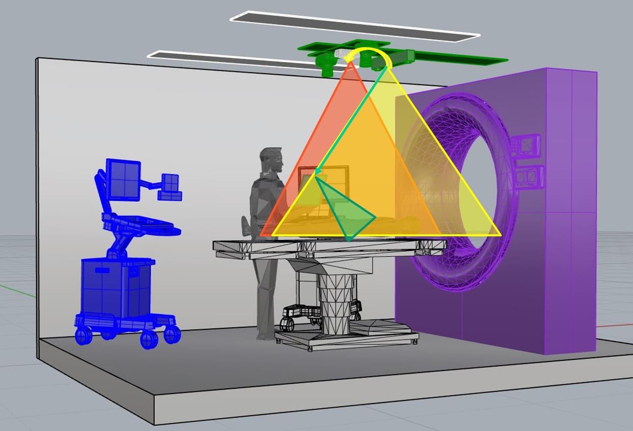
To combine the various acquisitions without permanently fixing the sensors to the CT device, the
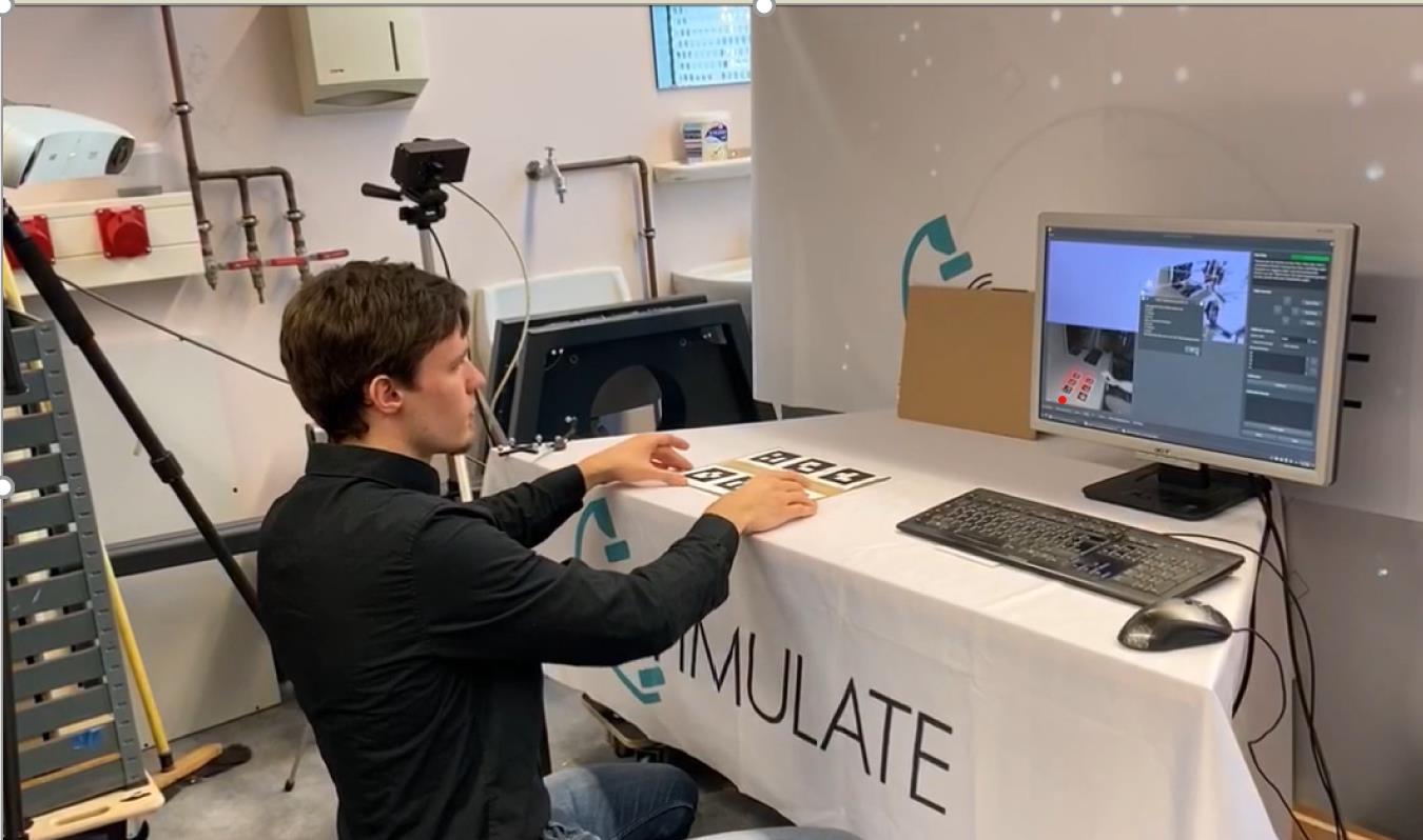
approach relies on an optical tracking system: marker-based detection is used to track the 3D
position and orientation of the sensor modules and the patient. A time-of-flight camera
installed above the CT table forms the center of the coordinate system, into which further –
including freely movable – devices are transformed. In addition, QR codes and, above all,
Bluetooth beacons proved effective in reliably detecting and identifying devices in the
examination room even when they are optically obscured.
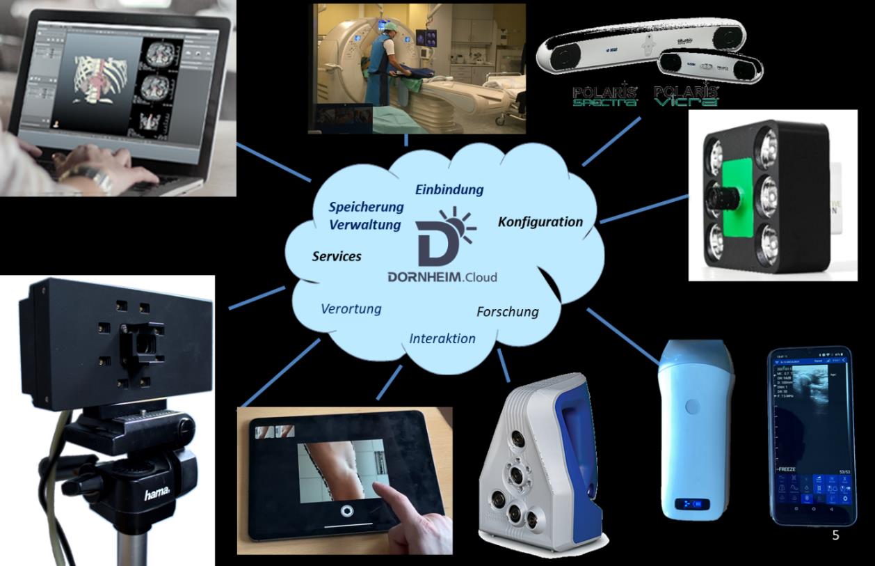
A web-based, adaptive user interface was developed for controlling and integrating the sensors.
A configuration file automatically embeds a device's available parameters into the web
interface; a template function allows preconfiguration by administrative users. Technically, the
concept builds on open standards (ROS) and a containerized infrastructure (Docker), in which the
web interface and device connection communicate bidirectionally via the WebSocket protocol – so
the system can be flexibly transferred to other devices and interfaces.
Use case: wound care
Wound care was chosen as an exemplary use case – a complex field shaped by intersectoral and
interdisciplinary work in which a wide variety of imaging devices is used. New mobile devices
with integrated 3D camera technology (iPhone Pro, iPad Pro) opened up the possibility of
capturing 3D scans, for example of wounds, directly with a handheld device. In prototype apps,
interaction concepts for touch interfaces were developed that allow structures to be marked and
their areas and dimensions to be calculated to scale – helpful for treatment and documentation.
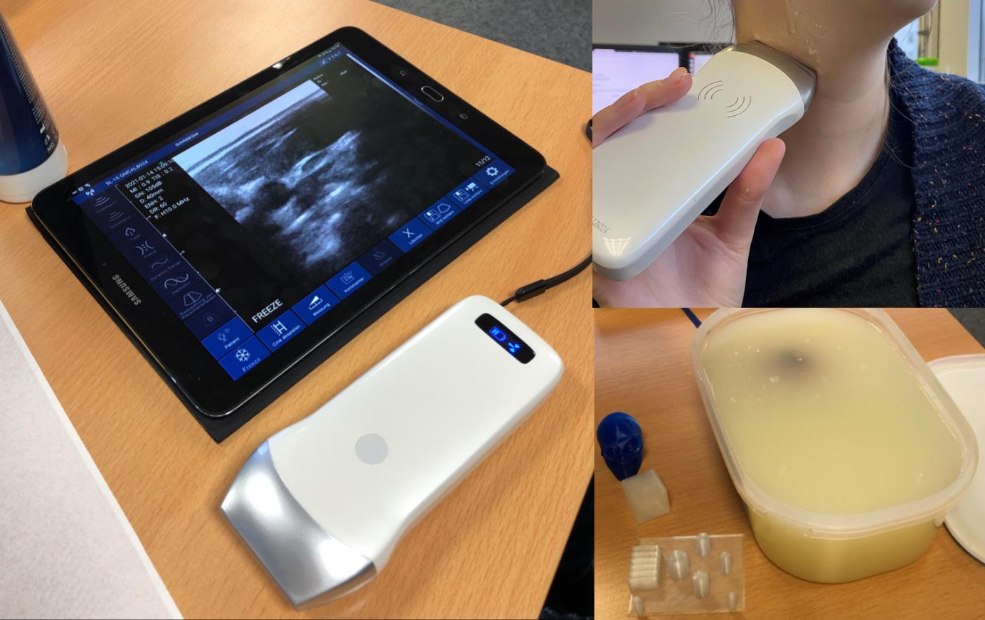
Beyond this, mobile, Wi-Fi-enabled ultrasound probes and a hyperspectral camera were integrated,
and their acquisitions were fused with 3D scans. Through the cloud-based system, multiple users
can view and discuss the results together across locations – an important building block for
telemedical and interdisciplinary collaboration.
Contributions by Dornheim Medical Images
Building on the Dornheim Segmenter®, matured over many years, Dornheim brought its expertise in
image analysis, 3D reconstruction, and visualization to the collaborative project. Among other
things, methods were developed for the spatial localization and calibration of the sensor
modules, for 3D surface reconstruction, for the multimodal visualization of overlaid volume
data, as well as cloud concepts for compute-intensive image processing and centralized data
management. The subproject was successfully completed from both a scientific and a technical
standpoint.