3D models from medical image data
created automatically and web-based
Illustrative three-dimensional models (3D models) derived from a patient's medical image data
could provide significant decision support in this regard – for both the physician and the
patient. This applies both to virtual 3D models, whose outer and inner shapes (morphologies)
can be easily examined interactively from all sides on a computer, and to the plastic 3D
models additively manufactured from them (e.g., via 3D printing).
Their production, however, requires many manual work steps that are carried out by in-house
experts or service providers in close coordination with the physician. Because of the
considerable amount of time this workflow entails, it is difficult to integrate into everyday
clinical practice.
In our development work we focus on prostate cancer. With a large number of factors that
influence the chances of recovery as well as the treatment options, it is a characteristic
use case and is therefore very well suited as an example, as well as for a later extension to
further clinical conditions.
Background information on the clinical condition and the motivation
Prostate cancer is the most common cancer among men and, after lung and colorectal cancer,
the third most common cancer-related cause of death in Germany. Many tumors are discovered by
chance, or grow so slowly that they never cause symptoms. Screening examinations also mean
that the majority of tumors are diagnosed at an early stage. Good chances of recovery are then
available. If the tumor has spread to other organs, however, the patient can usually only be
treated palliatively.
Besides the stage, the morphology of the tumor also has a major influence on the therapy and
the chances of survival. The spectrum of treatment options ranges from active surveillance to
chemotherapy, radiation therapy, or surgical removal, which entail severe side effects and
damage to surrounding organs. To avoid unnecessary damage, a very precise diagnosis as well as
therapy planning is crucial. The successes achieved in medical imaging encourage us in our
endeavor to make the generated 2D slice images more comprehensible, easier to evaluate, and
usable for more areas of application with the help of 3D representations and 3D models.
Our vision
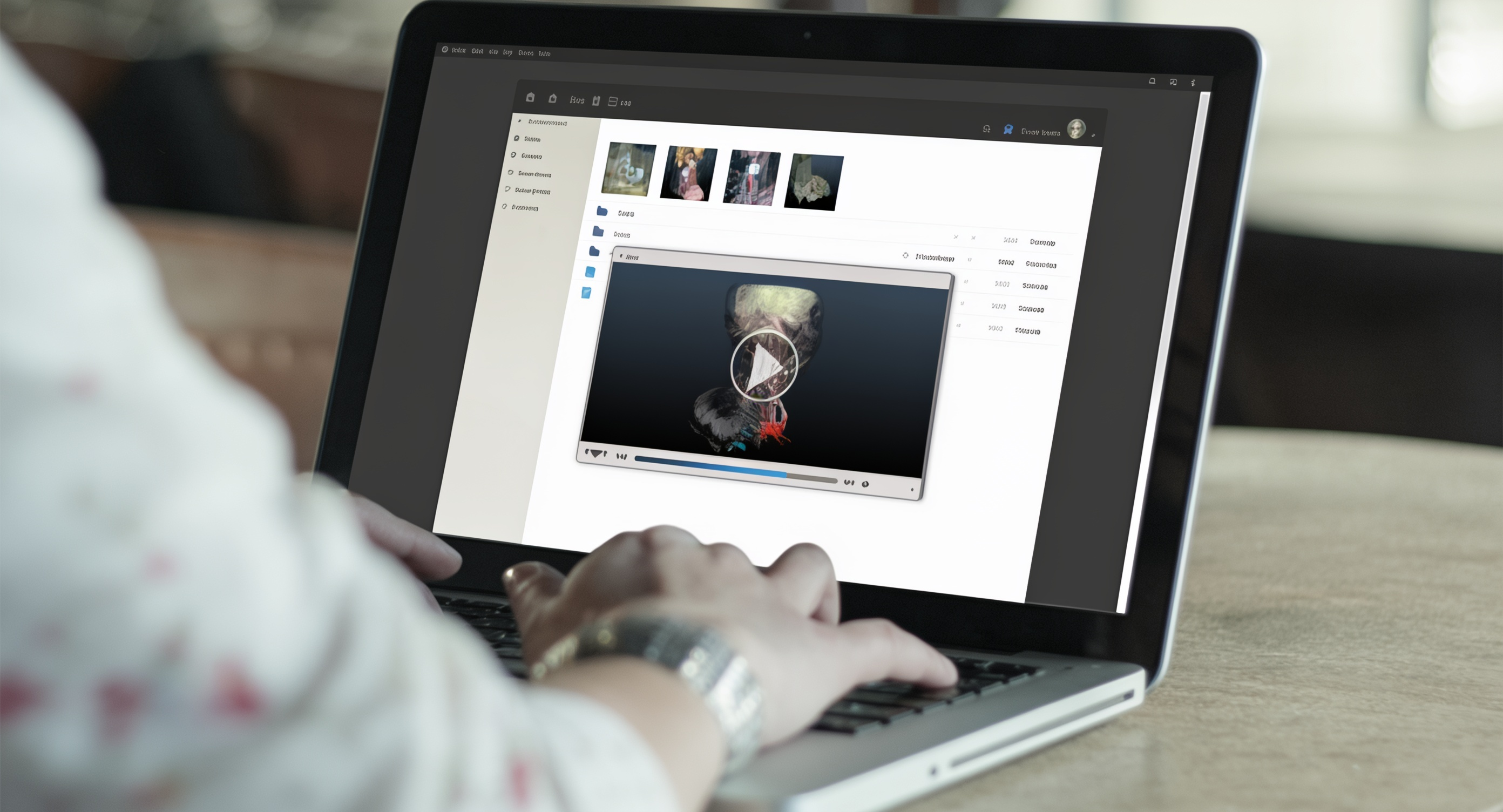
Our vision is to offer medical experts a platform through which model generation and
production can be delegated easily, internally or externally, and through which communication
is simplified. To this end, physicians should be able not only to upload their patients'
image data securely, but also to make it available to the experts who further process,
annotate, and in part even segment the data themselves – at any time and from various local
or mobile devices. As part of the research project, intelligent interaction techniques,
(partial) automation of image analysis and segmentation techniques, corresponding interfaces,
and further innovative online services are researched and developed.